
MBF Bioscience Solves Your Big Data Challenges
The ability to efficiently acquire large experimental data sets of 2D, 3D and 4D images is advancing rapidly across science. Tools

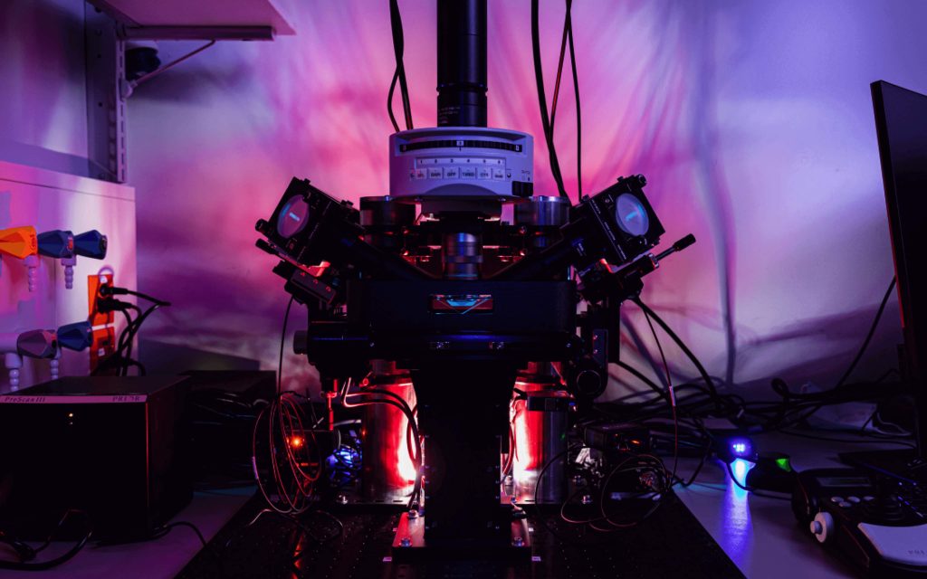
ClearScope is a revolutionary light sheet theta microscope system designed to quickly and easily image cleared specimens of nearly any size at subcellular resolution. Its patented technology has been developed through a collaboration between Dr. Raju Tomer at Columbia University and MBF Bioscience. ClearScope is designed to meet the unique challenges inherent to imaging cleared organs and brain specimens with high-resolution optics capable of distinguishing and obtaining quantifiable imaging data pertaining to cells (e.g. neurons, glial cells), vessels and subcellular structures like dendritic spines.
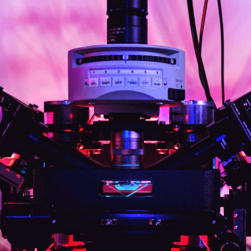
Versatile Light Sheet Imaging
ClearScope provides a unique combination of speed, 3D resolving power, tissue depth penetration, and low phototoxicity compared to confocal and multiphoton microscopy. The patented Light Sheet Theta Microscopy geometry is suitable for imaging a wide array of samples from embryos, to whole mouse brains up to large slabs of primate brain tissue. Virtually all clearing techniques are compatible with ClearScope via its Intelligent Refractive Index Compensation (IRIC). The modular hardware design allows for scanning of biologic structures at a variety of magnifications (4x-25x) and up to 7 laser wavelengths customizable to researcher needs.
Move Your Research Forward with Advanced Technology
ClearScope is your complete light sheet imaging solution designed with automation and efficiency in mind to help researchers increase scientific throughput without having to be an expert in optics, writing software or hardware control. For those users who want to experiment with imaging parameters and explore innovative ways of imaging cleared tissue specimens, the advanced user mode of ClearScope provides full control over all of the hardware components. A basic user mode for simplified operation includes automated light sheet optimization tools and streamlined workflows.
ClearScope has been developed with support from the National Institute of Mental Health (NIMH)
| Maximum Imaging Depth | 12mm (working distance depends on choice of objective lens) |
| Maximum Specimen Size | • 114mm x 75mm x 12mm with standard stage • 150mm x 100mm x 12mm with ext. travel stage |
| Refractive index range | 1.38 – 1.56 (i.e. compatible with CLARITY, CUBIC, Scale, iDisco, BABB, etc.) |
| Horizontal (XY) optical resolution | 0.325 μm (with 20x objective lens) |
| Laser wavelengths | 405 / 488 / 561 / 640 nm standard; (customizable up to seven lasers) |
| Specimen chambers | for whole mouse or rat brains, tissue slabs from primates and human specimens, plus custom chambers available for other size specimens |
| Single field-of-view pixel resolution | 2048 x 2048 pixels |
| Image digitization | 16 bit |
| Light sheet thickness | 2 - 6μm depending on optics |
| Peak quantum efficiency (QE) | 95% @ 560nm |
Detection Objective Lens:
| Magnification | Numerical Aperture | Working Distance (mm) |
| 4x | 0.28 | 10 |
| 10x | 0.60 | 8 |
| 17x | 0.40 | 12 |
| 20x | 1.00 | 8 |
| 24x | 0.70 | 10 |
| 25x | 0.95 | 8 |
BrightSlice is a powerful, all-in-one software solution designed specifically for light-sheet microscopy. From acquisition to analysis, handle teravoxel-scale imaging with unprecedented ease and precision.
Everything you need in one intuitive interface:
Generate publication-quality data with comprehensive processing tools:
Seamlessly connects with our professional analysis suite:
Plays well with your existing tools through OME-TIFF export:
Download ClearScope product sheet here.
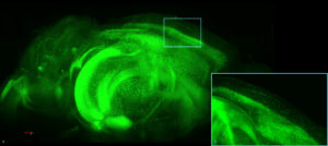
(A) 3D visualization maximum intensity projection of a transgenic mouse brain expressing eGFP under control of the Thy-1 promotor. The brain was cleared using Binaree Rapid Tissue Clearing (Binaree, Inc., Daegu, Republic of Korea; the 3D image was captured using a 10x magnification objective. (B) A Zoomed visualization of cortical neurons in the image.
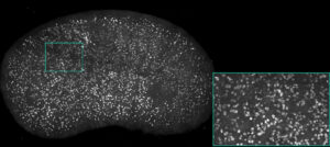
Maximum intensity projection of mouse kidney with vessels and glomeruli labeled using Lycopersicon Esculentum (Tomato) Lectin (LEL, TL), DyLight 649 (ThermoFisher Scientific, Waltham, MA, USA) (red). The kidney was cleared using Binaree Rapid Tissue Clearing (Binaree, Inc., Daegu, Republic of Korea). Image acquired at 10x magnification.
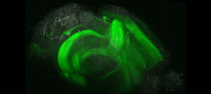
3D maximum intensity projection of a transgenic mouse brain expressing eGFP under control of the Thy-1 promotor (green), with vessels labeled using Lycopersicon Esculentum (Tomato) Lectin (LEL, TL), DyLight 649 (ThermoFisher Scientific, Waltham, MA, USA) (red). The brain was cleared using Binaree Passive Tissue Clearing (Binaree, Inc., Daegu, Republic of Korea); the 3D image was captured using a 10x 0.60 magnification objective.
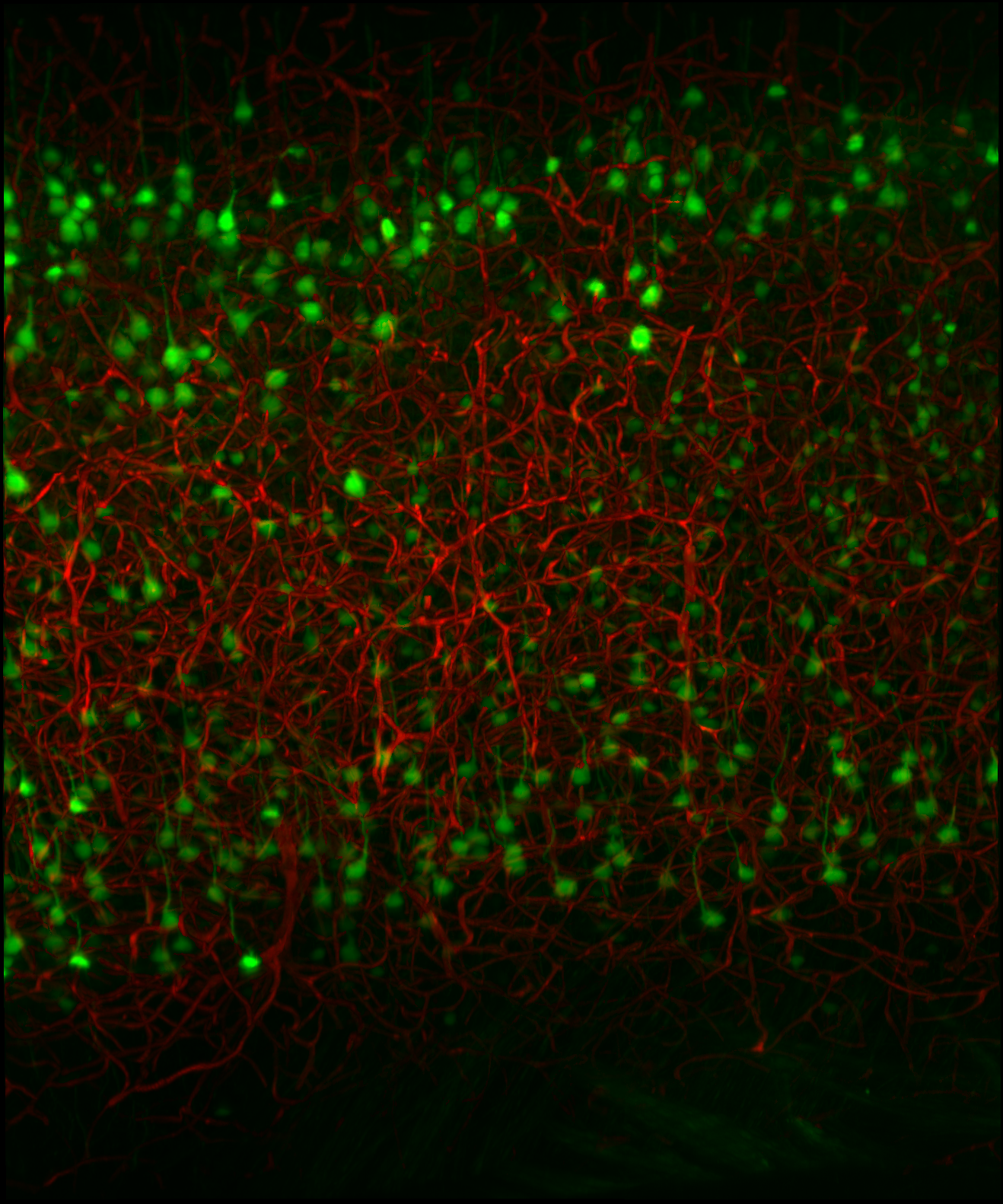
Maximum intensity projection of a portion of a 3D image of a transgenic mouse brain expressing eGFP under control of the Thy-1 promotor (green), with vessels labeled using Lycopersicon Esculentum (Tomato) Lectin (LEL, TL), DyLight 649 (ThermoFisher Scientific, Waltham, MA, USA) (red). The brain was cleared using Binaree Rapid Tissue Clearing (Binaree, Inc., Daegu, Republic of Korea; the 3D image was captured using a 10x 0.60 magnification objective.
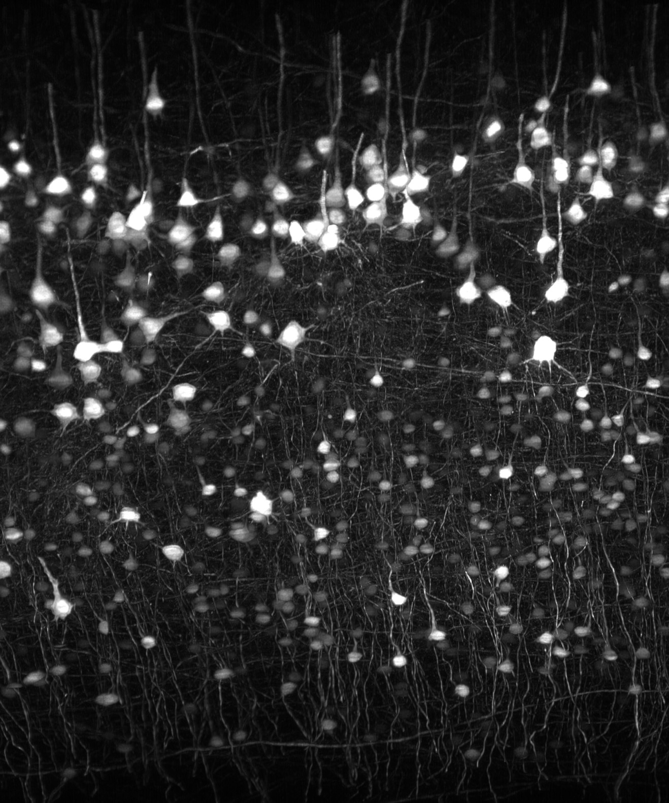
Image acquired on ClearScope and deconvolved using NeuroDeblur. Transgenic mouse brain expressing eGFP under control of the Thy-1 promotor show in a maximum intensity projection. The brain was cleared using Binaree Rapid Tissue Clearing (Binaree, Inc., Daegu, Republic of Korea); the 3D image was acquired at 10x magnification using 488 nm excitation lasers.
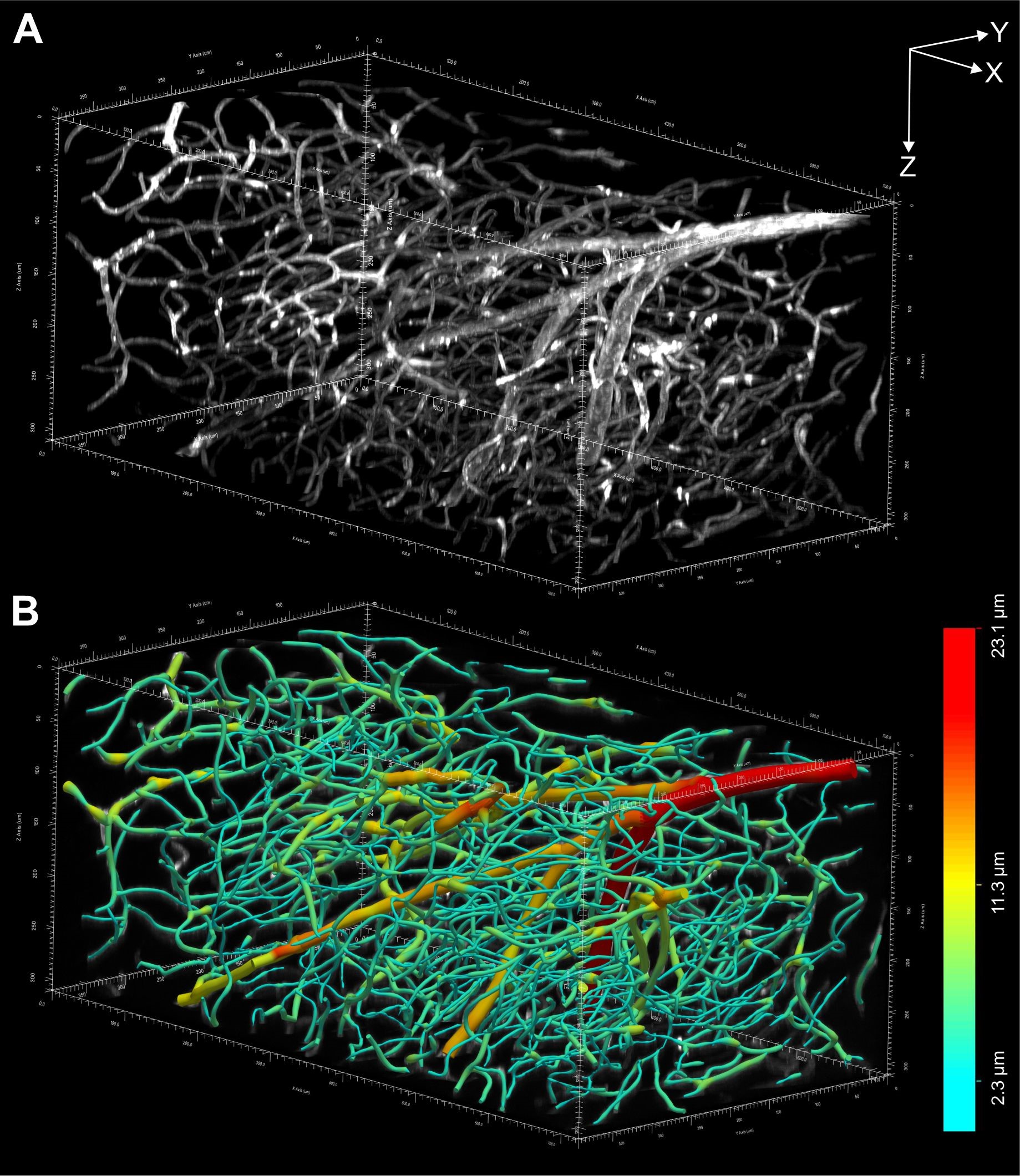
(A) 3D representation of the 3D image of an entire, intact mouse brain with vessels labeled using Lycopersicon Esculentum (Tomato) Lectin (LEL, TL), DyLight 649 (ThermoFisher Scientific) acquired using a ClearScope (B) Vessels in (A) digitally reconstructed for quantitative analysis using the automatic 3D reconstruction software, Neurolucida 360. The vessel diameters are color-coded as shown on the right. Each of the arrows in (A) represents 100 µm.
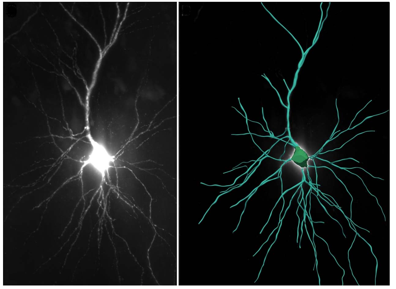
Left: A portion of a human postmortem entorhinal cortex cleared using iDisco8. Neurons were filled with biocytin dye following patch-clamping and subsequent staining with streptavidin tagged with Alexa 555. The 3D image was captured using a ClearScope. Right: A neuron from the left image, digitally reconstructed for quantitative analysis using the automatic 3D neuron reconstruction software, Neurolucida 360
ClearScope components are upgradeable and customizable to suit the imaging needs of researchers in a variety of fields. Researchers can choose the number of illumination lasers (up to seven), objective lenses, specimen holders, optical filters, and more, to maximize effectiveness and accommodate a wide range of both clearing methods and budgets. This makes ClearScope the most adaptable light sheet microscope system to fulfill all the needs of multiple research labs or core facilities.
ClearScope’s patented light sheet theta microscopy technology applies two light sheets, oblique to the specimen and detector axes to image tissue specimens of nearly any lateral size at high resolution, taking full advantage of the working distance of high NA detection objectives. The unique arrangement of the dual oblique illumination allows for gentle scanning of your large intact cleared tissue samples, mitigating redundant illumination of the scanned areas and resulting in more uniform excitation of the sample, thereby reducing imaging artifacts.
Video caption: Rapid volumetric calcium imaging of highly motile Hydra. Courtesy: Tomer Lab
Choosing the best clearing method for a given application is dependent on a number of factors including availability, efficiency, conservation of protein-based fluorescence, immunostaining compatibility, etc. ClearScope’s Intelligent Refractive Index Compensation (IRIC) automatically adjusts the depth of the light sheet as a function of refractive index of the tissue, which enables optimal imaging of cleared tissue specimens with various clearing protocols, e.g. Clarity, CUBIC, iDisco, etc. ClearScope supports a wide variety of detection objective lenses to address your specific application requirements.
Light-sheet microscopy generates extremely large data sets. ClearScope uses efficient data compression to minimize data storage requirements and maximize speed. We have developed cutting edge image processing solutions that dramatically speed up multi-TB image stitching, viewing and analysis. A highly versatile 3D visualization environment and a full suite of image optimization tools with state-of-the-art functionalities are included to support your analysis and publication needs.
ClearScope’s innovative design enables the use of high numerical aperture (NA) and long working distance objective lenses for better optical sectioning capability. Synchronized scanning of the light sheets with exposure of the rolling shutter readout of the sCMOS camera effectively creates a virtual slit, reducing out of focus light and improving signal to noise ratio. Simultaneous 2-axis scanning focuses the light sheet ensuring only the thinnest part of the light sheet intersects the detection plane and optimizes image quality across the field-of-view of the objective.
Read our white paper on how to streamline handling, processing, and analyzing extensive datasets from light sheet microscopes, allowing researchers to prioritize scientific discovery over data management challenges.

The ability to efficiently acquire large experimental data sets of 2D, 3D and 4D images is advancing rapidly across science. Tools

For Immediate Release Williston, VT (December 10, 2020) — The ability to image large, intact biological specimens is about to get
For Immediate Release: Williston, VT (February 04, 2020) — MBF Bioscience’s revolutionary light sheet microscope system, ClearScope, sets a new standard
MBF products are used across the globe by the most prestigious laboratories.


MBF’s products utility is underscored by the number of references it receives in the worlds most important scientific publications. See examples below:
Migliori, B., Datta, M. S., Dupre, C., Apak, M. C., Asano, S., Gao, R., … Tomer, R.
(2018). Light sheet theta microscopy for rapid high-resolution imaging of large biological samples. BMC Biology, 16(1). doi: 10.1186/s12915-018-0521-8View Publication

BMC Biology
ClearScope is compatible with a wide array of tissue clearing protocols (e.g. Clarity, CUBIC, iDisco, uDisco, SeeDB, Sca/e, etc…) using intelligent refractive index compensation (IRIC). ClearScope automatically adjusts the depth of the light sheet as a function of the refractive index and depth of the tissue sample, enabling a single detector objective to image specimens cleared with various clearing techniques. Users can select the appropriate configuration settings for their tissue sample easily within the intuitive GUI.
The ClearScope system supports a variety of specimen chambers as well as standard slide sizes. Chambers are specifically designed to accommodate the dual oblique light sheets and separate the tissue specimen from the immersion media while minimizing the required volume of costly clearing reagents. Chambers and slides are loaded into the immersion media reservoir using specimen holders that precisely locate the sample in a repeatable manner using a magnetic/mechanical detent, facilitating repeated imaging experiment capability. Customized chambers/holders are available for your specific applications.
The ClearScope software includes a highly versatile 3D visualization environment and a full suite of image optimization tools with state-of-the-art functionalities to support your analysis and publication needs. A quick preview mode enables you to visualize multi-TB image volumes immediately following acquisition. Advanced image stitching produces 3D image volumes suitable for quantitative analysis to produce accurate data for analyzing morphometry, cell populations, sub-cellular processes, vasculature and fluorescence properties.
At the core of the patented LSTM technology is the use of dual illumination pathways oblique to the specimen and detection pathway. This provides ClearScope with the ability to image thicker tissue specimens over a large lateral area (XY) at higher optical resolutions while maintaining fast imaging speed, high imaging quality, and low photo-bleaching. Two intersecting light sheets form an ultra-thin light sheet (2-6um) that optimizes axial image resolution and quality, produces uniform illumination and reduces out of focus features.
Optimized data storage systems with fast transfer rates take the stress out of working with multi-TB image datasets. MBF Bioscience offers high performance imaging workstations coupled to large capacity, expandable RAID networked attached storage units using 10GB/sec network connections for fast data transfer improving productivity. We can custom configure a network storage solution tailored to your lab's needs.
The axial resolution is dependent on the depth of field of the detection objective and the thickness of the light sheet used for imaging the sample. ClearScope can be configured with a variety of detection objectives, ranging from 4x/0.28NA to 25x/.95NA. The optical sectioning capability of lower NA objectives can be significantly improved by the thin light sheet excitation of the sample, whereas higher NA objectives are limited by the depth of field of the objective itself (which is inversely proportional to the square of the detection NA). In both cases, ClearScope synchronizes scanning of the thinnest part of the light sheet with the rolling shutter exposure of the camera to optimize image quality and improve the signal to noise ratio.
"I rarely have encountered a company so committed to support and troubleshooting as MBF."

Andrew Hardaway, Ph.D. Vanderbuilt University
"MBF Bioscience is extremely responsive to the needs of scientists and is genuinely interested in helping all of us in science do the best job we can."

Sigrid C. Veasey, MD University of Pennsylvania
"I am so happy to be a customer of your company. I always get great help related with your product or not. With the experienced members, you are the best team I've ever met. All of your staff are very kind and helpful. Thank you for your great help and support all the time."

Mazhar Özkan Marmara Üniversitesi Tıp Fakültesi, Turkey
"We’ve been very happy for many years with MBF products and the course of upgrades and improvements. Your service department is outstanding. I have gotten great help from the staff with the software and hardware."

William E. Armstrong, Ph.D. University of Tennessee
"Our experience with the MBF equipment and especially the MBF people has been outstanding. I cannot speak any higher about their professionalism and attention for our needs."

Bogdan A. Stoica, MD University of Maryland
"MBF provides excellent technical support and helps you to find the best technical tools for your research challenges on morphometry."

Wilma Van De Berg, Ph.D. VU University Medical Center - Neuroscience Campus Amsterdam
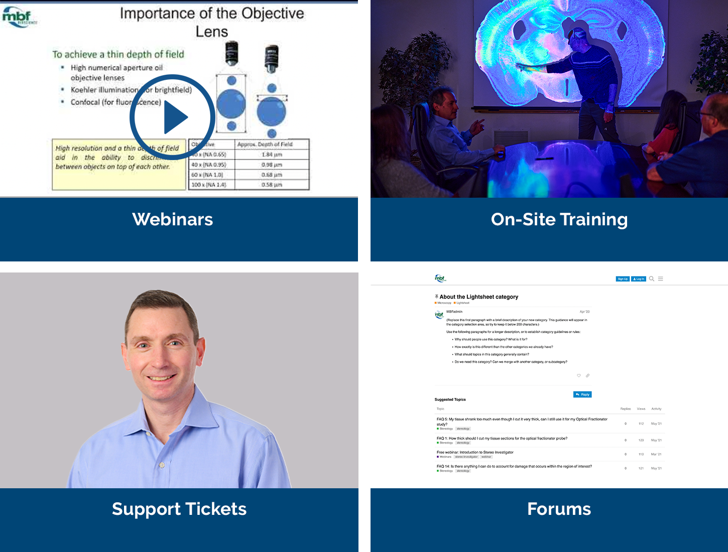
Our service sets us apart, with a team that includes Ph.D. neuroscientists, experts in microscopy, stereology, neuron reconstruction, and image processing. We’ve also developed a host of additional support services, including:
At MBF, we’ve spent decades understanding the needs of researchers and their labs — and have a suite of products and solutions that have been specifically designed for the needs of today’s most important and advanced labs. Our commitment to you is to spend time with you discussing the needs of your lab — so that we can make sure the solutions we provide for you are exactly what you’ll need. It’s part of our commitment to supporting you — before, during, and after you’ve made your decision. We look forward to talking with you!


