
Vesselucida Helps Researchers Quantify Post-Injury Capillary Damage and Regeneration
Our health depends on the ability of blood vessels to deliver nutrients and remove metabolic byproducts from organs and muscle systems.
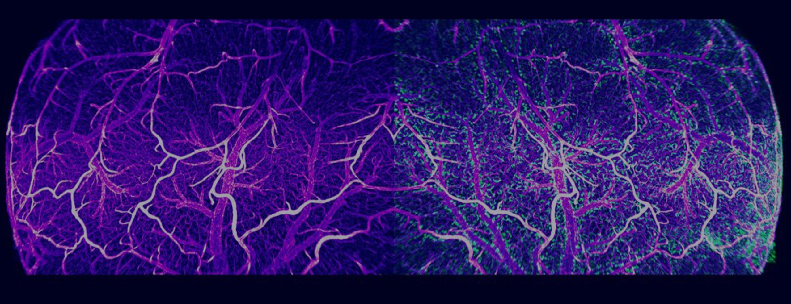
The Vesselucida system is uniquely designed to control your microscope hardware and provide purpose-built tools for accurate tracing of microvascular networks directly from histological specimens. Create reconstructions based on ground truth and use the wide array of built-in quantitative analysis tools to fuel your research in fields including cancer, diabetes, strokes and other conditions that affect microvasculature
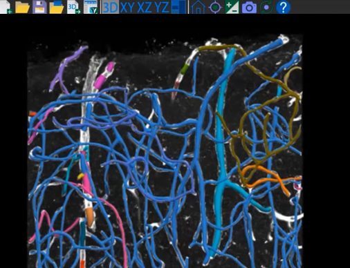
The Vesselucida system for vasculature reconstruction and quantitative morphometric anaysis is based on the ground truth—you trace directly from the specimen on the microscope or from previously captured images. The tracing tools are specialized for microvasculature, making it clear which tool to use and why; in addition, you can also edit traced structures as needed. Vesselucida works with a wide array of 2D and 3D images, even large 2D and 3D images of intact tissue specimens.
Vesselucida fully integrates with your microscope system hardware to control motorized stages, focus, cameras, objective changers, and optical filters. Whether you need a new microscope system or want to use Vesselucida with your existing microscope hardware, our technical sales specialists will work with you to understand your current and future research interests, and choose microscope components that will help you achieve your research goals and fit your budget.
|
Minimum System Requirements |
|
|
Operating System |
Windows 10, 64-bit |
|
Processor |
4-core |
|
CPU RAM |
16 GB |
|
GPU RAM |
>6 GB |
|
Recommended System Requirements |
|
|
Operating System |
Windows 10, 64-bit |
|
Processor |
8-core |
|
CPU RAM |
64 GB |
|
GPU RAM |
>8 GB |
|
Storage |
Solid state drive(s) |
|
|
*Certain high throughput Acquisition systems will require a NAS anywhere from 10 to 200TB in size |
|
Input Specifications |
|
|
Supported image file formats |
CZI, HDF5, H5, IMS, JP2, JPG, JPEG, JPF, MJC, JPX, LIF, LSM, ND2, OIB, OIF, TIF, TIFF, SVS |
|
Output Specifications |
|
|
Model data output formats |
XML*, ASC, DAT, SVG |
|
|
* Our XML data file format, the Neuromorphological File Specification (NFS), was recently endorsed as a standard by the INCF. |
|
Image file output formats |
JP2, JPX, TIFF |
|
Movie export format |
MP4 |
Supported Image File Formats: PDF
Vesselucida Helps Researchers Quantify Post-Injury Capillary Damage and Regeneration
>> Learn More
New Therapy May Aid Heart Repair After Heart Attack
>> Learn More
Scientists use Vesselucida 360 to quantify brain vasculature in mTBI model
>> Learn More
Download Vesselucida product sheet here
Vesselucida can serve as your microscope image-acquisition system. With features for acquisition of high-quality images and z- stacks, your system can be used for much more than vessel tracing. You can also capture high-quality 2D and 3D whole-slide images (high resolution digital montages of your specimen) with the addition of the Slide-Scanning Module. Use image processing features such as background correction to obtain clean, even, image quality. Vesselucida can acquire a wide array of images, using brightfield and multi-channel fluorescence that surpass the quality of most slide scanners in a box.
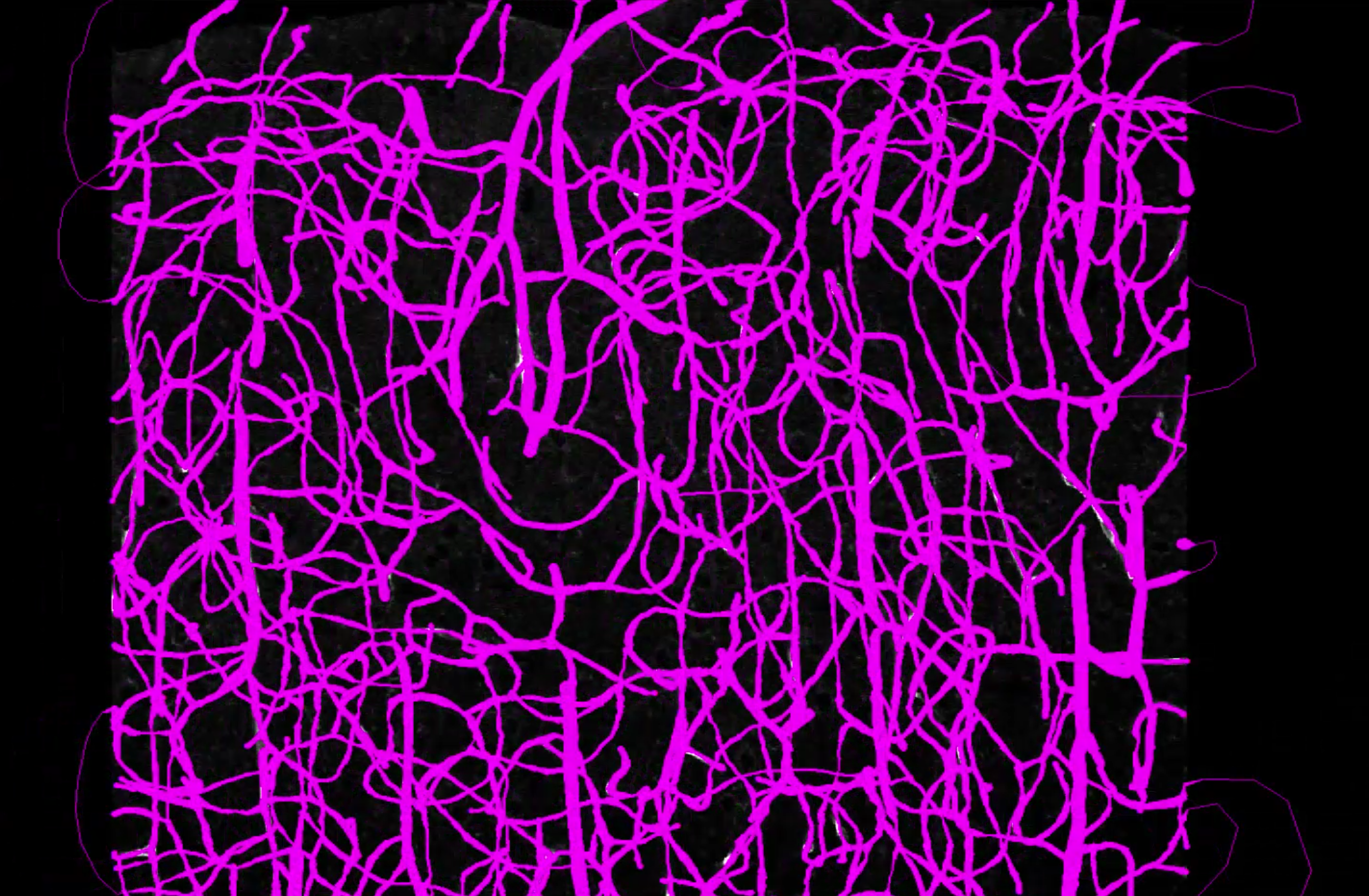
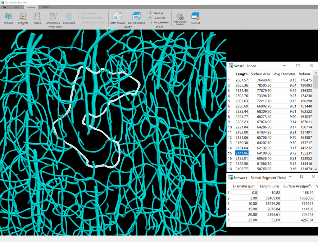
Our rigorously tested analyses provide accurate and robust results that you and others can trust for publications. Use Vesselucida Explorer, the companion analysis software, to perform sophisticated analyses that help answer your research questions.
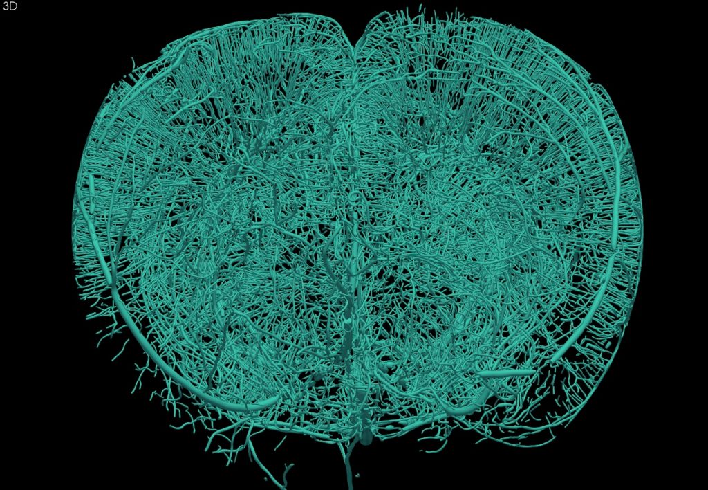
Vesselucida Explorer provides vasculature-specific metrics such as segment and node counts, frequency of anastomoses, vessel surface and volume, and more.
Vesselucida also supports importing image stacks and whole-slide images acquired using other systems. Working with whole-slide images enables you to create vasculature reconstructions and analyze them on any computer, even those not connected to a microscope.
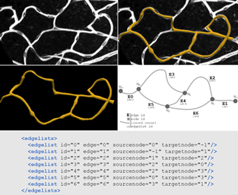
We support open science through data sharing, accessibility, integrity, and reproducibility. The published MBF Bioscience digital reconstruction data file format, the Neuromorphological File Specification (NFS), was recently endorsed as a standard by the INCF.
The data elements in this NFS format were implemented specifically to ensure that files produced in Vesselucida are findable, accessible, interoperable, and reusable (FAIR). Vesselucida embraces these data standards and provides microscopy image and experimental data provenance to enhance the ease of repurposing data generated with the software. Encoded in the well-recognized and readable XML format, the modeling elements specify anatomical information in a calibrated 3D coordinate system with appropriate units. To learn more about the key elements of the file format and its relevant structural advantages, read our publication.
Vesselucida is used across the globe by the most prestigious laboratories.


Our health depends on the ability of blood vessels to deliver nutrients and remove metabolic byproducts from organs and muscle systems.
Vesselucida’s utility is underscored by the number of references it receives in the worlds most important scientific publications.
Alese, Lassig et al.
Characterization of cutaneous microvasculature with 3D imaging: a feasibility study in a cohort of head and neck surgery patients with attention to smoking statusView Publication
Buncha, V., K. A. Fopiano, et al.
Mice deficient in endothelial cell-selective adhesion molecule develop left ventricle diastolic dysfunctionView Publication

Brady, E. L., O. Prado, et al.
Engineered tissue vascularization and engraftment depends on host modelView Publication

Jacobsen, N. L., C. E. Norton, et al.
Myofibre injury induces capillary disruption and regeneration of disorganized microvascular networksView Publication

Gama Sosa, M. A., R. De Gasperi, et al.
Late chronic local inflammation, synaptic alterations, vascular remodeling and arteriovenous malformations in the brains of male rats exposed to repetitive low-level blast overpressuresView Publication

Sullivan, A.E., Tappan, S.J., Angstman, P.J. et al.
A Comprehensive, FAIR File Format for Neuroanatomical Structure Modeling. Neuroinform (2021). https://doi.org/10.1007/s12021-021-09530-xView Publication

Gama Sosa, M.A., De Gasperi, R., Pryor, D. et al.
Low-level blast exposure induces chronic vascular remodeling, perivascular astrocytic degeneration and vascular-associated neuroinflammation. acta neuropathol commun 9, 167 (2021). https://doi.org/10.1186/s40478-021-01269-5View Publication

Button, E.B., Boyce, G.K., Wilkinson, A. et al.
ApoA-I deficiency increases cortical amyloid deposition, cerebral amyloid angiopathy, cortical and hippocampal astrogliosis, and amyloid-associated astrocyte reactivity in APP/PS1 mice. Alz Res Therapy 11, 44 (2019).View Publication

Yao, Y., Taub, A.B., LeSauter, J. et al.
Identification of the suprachiasmatic nucleus venous portal system in the mammalian brain. Nat Commun 12, 5643 (2021)View Publication

Editions of Vesselucida are available that work with or without a microscope. If your vessels span multiple sections or dozens of fields of view, you may find it quicker and easier to reconstruct vascular systems with a microscope, using Vesselucida – Microscope Edition. If you’re reconstructing vasculature that is relatively easy to image, you may benefit from the automatic reconstruction features available in Vesselucida 360.
Vesselucida systems can be integrated with hardware from all four major microscope manufacturers (Zeiss, Olympus, Leica and Nikon). Depending on your microscope model, components, and experimental needs, your current microscope system may require hardware upgrades. Some of the most important components are stage, focus drive, cameras, and light sources. Contact us to assess your microscope system and help you determine a configuration that facilitates your research goals and fits your budget.
Vesselucida is ideal for reconstructing vasculature in tissue specimen slides directly from a research microscope. Vesselucida also enables you to map and reconstruct anatomical regions through serial sections to create 3D models. You can acquire 3D image stacks and 2D or 3D scans of slides for publication or archival or image analysis away from the microscope.
Every license of Vesselucida and Vesselucida 360 includes access to Vesselucida Explorer companion software. This data-analysis tool can quickly and easily export your data to Excel or to the clipboard. It even includes batch exporting so that you can process hundreds of data files with just a few clicks!
No, you will be given access to a free copy of Vesselucida Explorer with every copy of Vesselucida or Vesselucida 360 you purchase. If you don’t own Vesselucida or you need more licenses for use on analysis-only computers, you can purchase stand-alone Vesselucida Explorer licenses.
Vesselucida 360 can automatically or semi-automatically reconstruct vasculature and anatomies from images and image stacks acquired using a variety of microscopy types. In contrast, Vesselucida includes microscope-control capability and manual tracing tools to reconstruct vessels in tissue specimen slides directly from a research microscope.
"Vesselucida 360 is an innovative tool for the characterization of cutaneous vasculature; using this tool, an increase in isolated elements was found to be associated with poorer wound healing outcomes ... Vesselucida 360 is a useful tool in objectively characterizing cutaneous microvascular structure via confocal microscopic tissue evaluation."

Alese, Lassig et al. JPRAS Open, 2025
"I rarely have encountered a company so committed to support and troubleshooting as MBF."

Andrew Hardaway, Ph.D. Vanderbuilt University
"Vesselucida enables us to visualize and quantify microvascular networks in a way that has not been done before. The ability to render 3D maps is integral to analysis of network structure, remodeling and blood flow distribution."

Dr. Nicole Jacobsen University of Missouri
"MBF Bioscience is extremely responsive to the needs of scientists and is genuinely interested in helping all of us in science do the best job we can."
Sigrid C. Veasey, MD University of Pennsylvania
"After examining different vessel quantification tools for use with neovessel formation in the heart, we chose to use Vesselucida 360 because it offered us flexibility in viewing and adjusting vessel geometries in 3D to accurately represent our microCT dataset. Also, the quantitative parameter extrapolation is excellent."

Dr. Kareen Coulombe Brown University
"I am so happy to be a customer of your company. I always get great help related with your product or not. With the experienced members, you are the best team I've ever met. All of your staff are very kind and helpful. Thank you for your great help and support all the time."

Mazhar Özkan Marmara Üniversitesi Tıp Fakültesi, Turkey
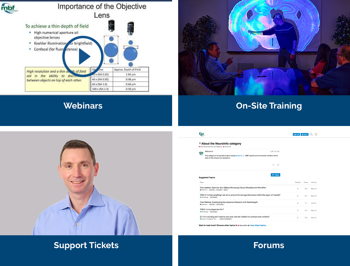
Our service sets us apart, with a team that includes Ph.D. neuroscientists, experts in microscopy, stereology, neuron reconstruction, and image processing. We’ve also developed a host of additional support services, including:
We offer a free expert demonstration of Vesselucida. During your demonstration you’ll also have the opportunity to talk to us about your hardware, software, or experimental design questions with our team of Ph.D. neuroscientists and experts in microscopy, neuron tracing, and image processing.


